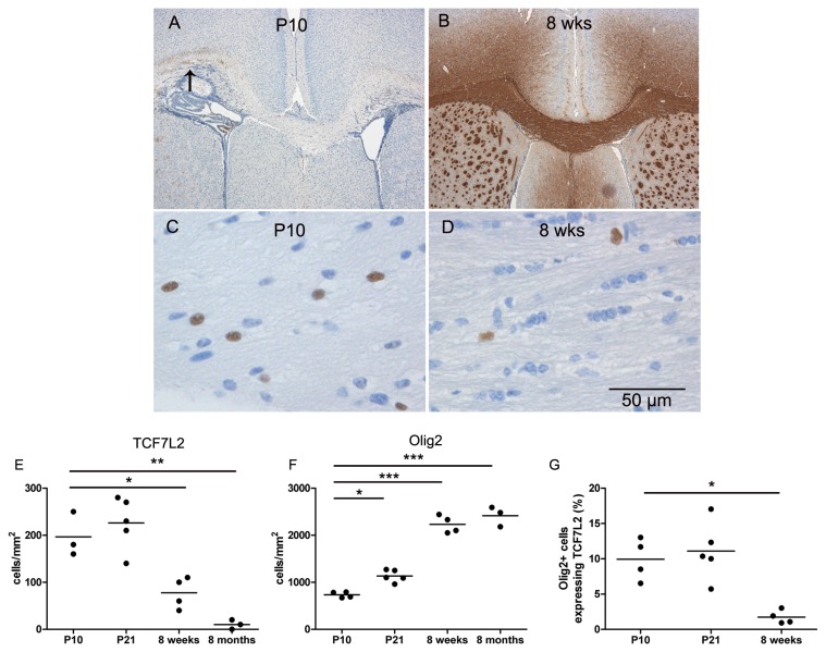
Figure 1. Expression of TCF7L2 during myelination in the CNS of mice.
At P10 single myelinated axons are observed in the lateral parts of the corpus callosum (arrow) (A) whereas in adult mice myelination is complete (B). High numbers of TCF7L2 expressing cells were observed in P10 mice (C), but only few TCF7L2-positive cells were found in adult mice (D) as quantified in the diagrams (E and F). The percentage of OLIG2 positive cells expressing TCF7L2 decreased significantly in adult mice (G).

