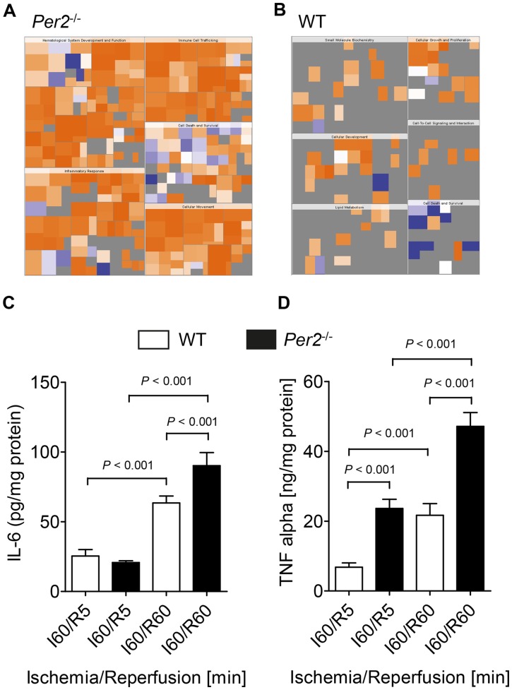
Figure 6. Initiation of a pro- inflammatory program in Per2 − /− mice during ischemia and reperfusion.
(A,B) Pattern recognition analysis (heat map of biological functions) from genes only regulated in Per2−/− (A) or WT (B) mice after 30 minutes of ischemia and 60 minutes of reperfusion. (C, D) Wildtype or Per2−/− mice were exposed to 60 minutes of ischemia and 5 (I60/R5) or 60 (I60/R60) minutes of reperfusion. The area at risk was excised and analyzed for IL-6 (C) or TNF-α (D) cardiac tissue concentration; n = 3 mice in all groups.

