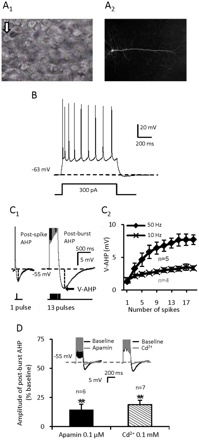
Figure 1. Identification of mPFC pyramidal cells and features of post-spike/burst AHP.

(A) A pyramidal cell in layer V/VI of the mPFC with a prominent apical dendrite. (B) A mPFC pyramidal cell which shows spike-frequency adaptation. (C) C1: The post-spike AHP and post-burst AHP with 13 spikes. Each inward depolarizing pulse is 2.8 nA for 2 ms. C2: The more the no. of depolarizing pulses are and the higher the depolarizing frequency is, the bigger the amplitude of post-burst AHP is. (D) The induced post-burst AHP is apamin sensitive and calcium dependent (**p<0.01 for Apamin vs. baseline; **p<0.01 for Cd2+ vs. baseline, paired t-test). Values are represented as mean ± SE.
