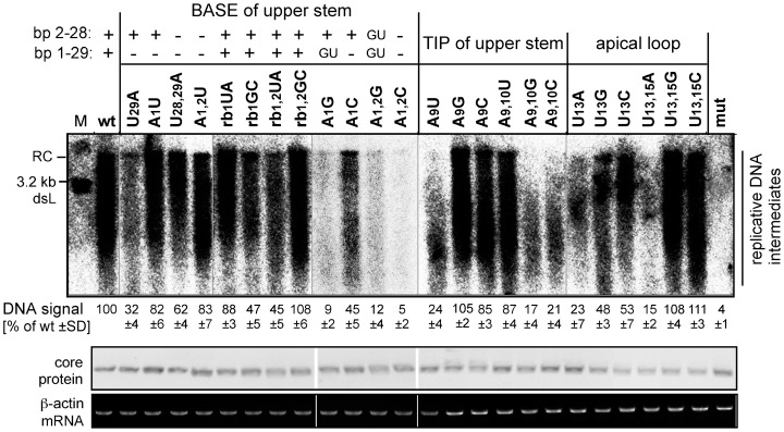
Figure 3. Impact of individual ε mutations on viral DNA accumulation.
HepG2 cells were transfected with the wild-type (wt) HBV expression vector pCH-9/3091, or derivatives containing the mutant 5′ ε sequences shown in Fig. 2B. The + or − signs indicate whether canonical base-pairs could form between residues at the A1-U29 and A2-U28 positions; potential G-U pairs are separately indicated. Viral DNAs from cytoplasmic nucleocapsids were monitored by Southern blotting (top panel) using a 32P-labeled HBV DNA probe; M, 3.2 kb restriction fragment corresponding to a unit length double-stranded linear (dsL) HBV genome. As controls, core protein and β-actin mRNA levels in the source lysates were monitored by Western blotting (middle panel) and RT-PCR (lower panel). Numbers below each lane show the accumulation of viral DNA replicative intermediates, measured by phosphorimaging, relative to those produced by the wild-type HBV construct which was set to 100. Mean values ± standard deviation were derived from two independent experiments.

