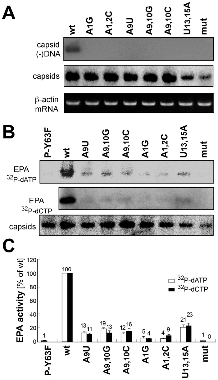
Figure 4. Direct confirmation of low DNA content in intact capsids derived from mutant ε constructs.
(A) DNA detection by molecular hybridization. Cytoplasmic capsids from cells transfected with the indicated constructs were separated by NAGE. After blotting, HBV DNA in the capsids was monitored by hybridization with a minus-strand specific probe, and capsids by immunodetection (panel labeled capsids). β-actin mRNA as determined by RT-PCR (panel labeled β-actin) served as loading control. (B) Endogenous polymerase assays (EPAs). One aliquot each of cytoplasmic capsids was subjected to EPA conditions in the presence of α-32P-dATP or α-32P-dCTP, then separated by NAGE. Labeled products associated with the capsids were visualized by autoradiography. Y63F refers to a replication-defective HBV construct in which the priming Tyr63 residue of P was replaced by Phe. A third aliquot from each sample was used for immunodetection of NAGE-separated capsids (panel labeled capsids). (C) Relative EPA activities. The bar graph shows the signal intensities generated by individual mutants for α-32P-dCTP and α-32P-dATP EPAs relative to that produced by wild-type HBV which was set at 100. Numbers are mean values from at least two independent experiments; error bars indicate standard deviation.

