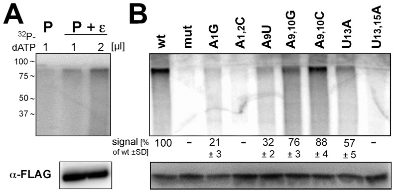
Figure 6. In vitro priming activities of selected ε RNAs showing reduced DNA accumulation in cells.
(A) Increased priming signals by co-expression of wild-type ε RNA and increased α-32P-dATP concentration. FLAG-tagged HBV P protein from cells transfected with only the P protein vector (lane P), or cotransfected with a wild-type ε RNA expression vector (lanes P + ε) was immobilized on anti-FLAG antibody beads. One third each of the immunoprecipitate was incubated with 1 µl or 2 µl of α-32P-dATP (3,000 Ci/mmol) as reported [44]; subsequently, the beads were boiled in SDS-PAGE sample buffer, and the released material was analyzed by SDS-PAGE and autoradiography. The remaining one third of the immunopellet was analyzed for FLAG-tagged P protein by Western blotting (panel anti-FLAG). (B) In vitro priming activities of selected ε RNA variants. FLAG-tagged P protein complexes with the indicated ε RNAs were expressed, affinity purified and subjected to in vitro priming conditions as in (A), using 2 µl α-32P-dATP. The molecular mass marker positions indicated on the left (in kDa) are approximations inferred from the respective marker protein positions on the SDS-PAGE gels used for the anti-FLAG immunoblots which were run in parallel under identical conditions. Numbers below the autoradiogram indicate mean signal intensities ± standard deviation from two independent experiments relative to that produced by the wild-type ε RNA complex which was set to 100.

