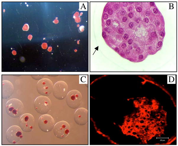
Figure 5.
Direct light microscopy photos with DTZ (A, 40×) and H&E (B, 400×) staining of the retrieved capsules at 130 days after transplantation. The arrow indicates the border of the capsule. DTZ (C, 40×) and insulin fluorescent stained (D, Confocal Microscopy) pictures of the retrieved capsules at day 329 after transplantation.

