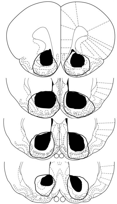
Figure 1. Histological results.
A: Smallest (dark) and largest (outline) lesion extents at each of four coronal planes, 2.70mm, 1.70mm, 1.20 mm, and 0.70 mm anterior to bregma (top to bottom). Section outlines are from Paxinos and Watson (1998) and are used by permission of Elsevier.

