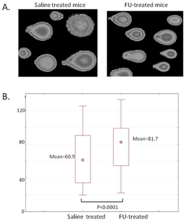
Figure 3.
Evaluation of DNA damage in the murine brain cells by alkaline Comet assay. Panel A: Epifluorescence microscopy images of the Comet assay slides after single-cell electrophoresis in alkaline conditions. Panel B: Quantitative analysis of images shown in Panel A using CometScore software (TriTek, Sumerduck, VA), as described in Materials and Methods section.

