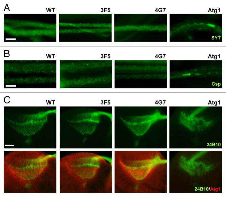
Figure 4. Axonal growth, projection and transport are unaffected by Fip200 loss. (A and B) Motor neurons from wandering-stage third instar larvae were immunostained with the indicated antibodies. In Fip200-null mutant (3F5 and 4G7) motor neurons, axonal traffic of synaptotagmin (SYT), (A) and cysteine string protein (Csp), (B) cargo molecules were normal and indistinguishable from WT motor neurons. However, the proteins became aggregated in Atg1-null (Atg1Δ3d/Df(3)BSC10) motor neurons. (C) Wandering-stage third instar larval brain was immunostained with the indicated antibodies. In both WT and Fip200-null mutant brains, axons of eye photoreceptor cells, visualized by 24B10 monoclonal antibodies, show stereotyped projection to lamina and medulla region, and Atg1 prominently localizes to axons in lamina of the brain optic lobes. Atg1-null (Atg1Δ3d/Df(3)BSC10) brains show disruption of this structure. Scale bars: 20 μm.

An official website of the United States government
Here's how you know
Official websites use .gov
A
.gov website belongs to an official
government organization in the United States.
Secure .gov websites use HTTPS
A lock (
) or https:// means you've safely
connected to the .gov website. Share sensitive
information only on official, secure websites.
