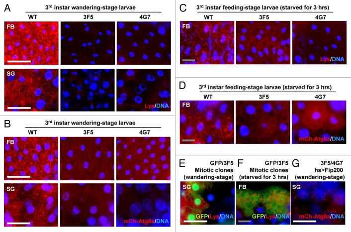
Figure 5. Fip200 is essential for developmental and starvation-induced autophagy. (A) Fat bodies (FB) and salivary glands (SG) of wandering-stage third instar larvae of WT, 3F5 and 4G7 flies were subjected to LysoTracker Red (Lys) and Hoechst 33258 (DNA) staining. (B) FB and SG of wandering-stage third instar larvae of hs > mCherry-Atg8a/+ (WT), hs > mCherry-Atg8a Fip2003F5/Fip2003F5 (3F5) and hs > mCherry-Atg8a Fip2004G7/Fip2004G7 (4G7) flies, heat-shocked at 37°C for 1 h to induce mCherry-Atg8a and recovered at 25°C for 3 h, were stained with Hoechst 33258 (DNA) and observed under fluorescence microscopy. (C) FB from feeding-stage third instar larvae of WT, 3F5 and 4G7, starved for 3 h, were subjected to Lys and DNA staining. (D) FB from feeding-stage third instar larvae of hs > mCherry-Atg8a/+ (WT), hs > mCherry-Atg8a Fip2003F5/Fip2003F5 (3F5) and hs > mCherry-Atg8a Fip2004G7/Fip2004G7 (4G7) flies, heat-shocked at 37°C for 1 h and incubated on 20% sucrose solution at 25°C for 3 h, were subjected to DNA staining. (E) SG from wandering-stage third instar larvae of y w hs-FLP; FRT82B Ubi-eGFP/FRT82B Fip2003F5 (GFP/3F5) flies were subjected to Lys and DNA staining. Absence of GFP marks Fip200-deficient cells, which also have less Lys staining. (F) FB from feeding-stage third instar larvae of y w hs-FLP; FRT82B Ubi-eGFP/FRT82B Fip2003F5 (GFP/3F5) flies, starved for 3 h, were subjected to Lys and DNA staining. (G) SG of wandering-stage third instar larvae of UAS-Fip200/+; hs > mCherry-Atg8a Fip2003F5/Fip2004G7 (3F5/4G7 hs > Fip200) flies, heat-shocked at 37°C for 1 h and recovered at 25°C for 3 h, were subjected to DNA staining. Scale bar: 200 μm (white), 50 μm (gray).

An official website of the United States government
Here's how you know
Official websites use .gov
A
.gov website belongs to an official
government organization in the United States.
Secure .gov websites use HTTPS
A lock (
) or https:// means you've safely
connected to the .gov website. Share sensitive
information only on official, secure websites.
