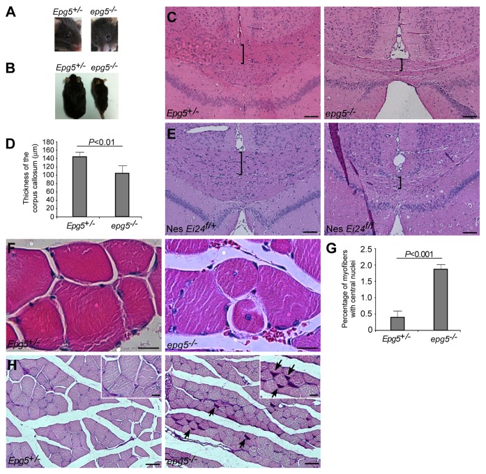
Figure 1. Neurological and muscular defects in epg5−/− mice. (A) epg5−/− mice do not show obvious cataracts. (B) epg5−/− mice have the same fur color as controls. (C) H&E staining of cerebra shows decreased thickness of the corpus callosum (brackets) in epg5−/− mice. Scale bar: 100 µm. (D) The thickness of the corpus callosum in mutant and control Epg5 mice. Mean ± SEM of 5 mice is shown. (E) H&E staining of cerebra shows decreased thickness of the corpus callosum (brackets) in Ei24flox/flox; nestin-Cre mice. Scale bar: 100 µm. (F) H&E staining of gastrocnemius muscles shows muscle atrophy and centrally nucleated fibers in epg5−/− mice. Scale bar: 10 µm. (G) The percentages of myofibers with central nuclei. Mean ± SEM of 3 mice is shown. (H) PAS staining of gastrocnemius muscles shows glycogen accumulation (arrows) in epg5−/− mice. Scale bar: 50 µm in main panels, 20 µm in insets.

An official website of the United States government
Here's how you know
Official websites use .gov
A
.gov website belongs to an official
government organization in the United States.
Secure .gov websites use HTTPS
A lock (
) or https:// means you've safely
connected to the .gov website. Share sensitive
information only on official, secure websites.
