
Figure 1. Huntingtin expression in cultured WT and BACHD astrocytes.
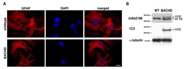
(A) GFAP immunocytochemistry on purified WT and BACHD astrocytes, whose nuclei are counterstained with DAPI. Both WT and BACHD cultured astrocytes show similar GFAP stain. Scale bar, 20μm. (B) Western blot analysis of protein extracted from cultured astrocytes with the monoclonal antibody 2166 (top panel) that stains endogenous mouse htt and full-length human mhtt in BACHD astrocytes. Only the endogenous mouse protein is found in WT mice, while both endogenous and full-length human mhtt are seen in the BACHD lane. The monoclonal antibody 1C2 (middle panel) recognizes an expanded polyQ epitope and reveals full-length human mhtt in BACHD astrocytes and no bands in WT. Immunoreactivity of α-tubulin was used as a control for gel loading (bottom panel).