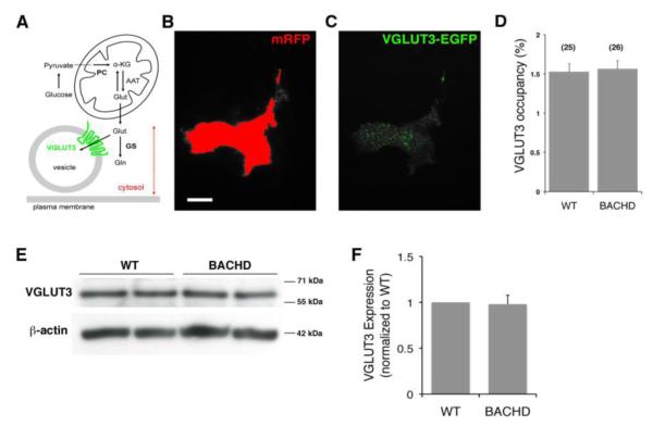
Figure 3. Vesicular glutamate transporter 3 (VGLUT3) levels are unaltered in BACHD astrocytes.
(A) Schematic representing regulation of glutamate in exocytotic glutamate release from astrocytes. Glutamate (Glut) can be synthesized in astrocytes de novo from glucose entry through the tricarboxylic acid cycle via pyruvate carboxylase (PC); glucose is broken down to pyruvate in the cytosol. Glutamate is converted from the cycle intermediate, α-ketoglutarate (α-KG), usually by transamination of aspartate via mitochondrial aspartate amino transferase (AAT). The synthesized glutamate once in the cytosol can then be converted to glutamine (Gln) by glutamine synthetase (GS), or transported into vesicles via vesicular glutamate transporters (VGLUTs), especially isoform 3 (VGLUT3). Drawing is not to scale. Dotted double arrow denotes intracellular penetration depth of total internal refection fluorescence (TIRF) illumination (~ 80 nm from the glass coverslip) used in experiments in B-D. (B-C) TIRF images of a WT astrocyte co-expressing mRFP (B) and VGLUT3-EGFP (C). (B) To designate the area within the TIRF field of an individual astrocyte, a threshold was applied to mRFP fluorescence emission (red). (C) This area was transposed on the VGLUT3-EGFP image, and VGLUT3-EGFP positive coverage was recorded (green). Scale bar in B, 20 μm. (D) The proportion of the cell area occupied by VGLUT3-laden vesicles (%) was similar in WT and BACHD astrocytes. Bars represent means ± SEMs of measurements from individual solitary astrocytes (numbers listed in parentheses). (E) Representative Western blots probing VGLUT3 from WT and BACHD astrocytes. Immunoreactivity of β-actin was used as a control for gel loading (bottom panel). (F) Quantification of Western blots shows no significant change in the level of VGLUT3 levels between WT and BACHD (n=3).

