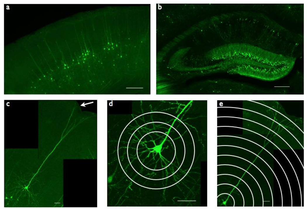
Figure 1. Thy1-GFP/Mreporter imaging strategy.
(a) Somatosensory cortex layer V pyramidal neurons are labeled with GFP throughout the cell body and dendritic tree in Thy1-GFP/M reporter mice. Scale bar = 250 µm.
(b) Hippocampus CA1 pyramidal neurons are labeled with GFP throughout the cell body and dendritic tree in Thy1-GFP/M reporter mice. Scale bar = 250 µm.
(c) Somatosensory cortex layer V pyramidal neurons extend their apical dendrites to the pial surface (arrow) and their basal dendrites deep into layer VI. Scale bar = 50 µm.
(d) Basal dendrites of somatosensory cortex layer V pyramidal neurons, quantified by Sholl analysis, measuring number of intersections between concentric circles drawn around the cell soma (Sholl radii) and dendritic branches. Scale bar = 50 µm.
(e) Apical dendrites of somatosensory cortex layer V pyramidal neurons, quantified by Sholl analysis. Scale bar = 50 µm.

