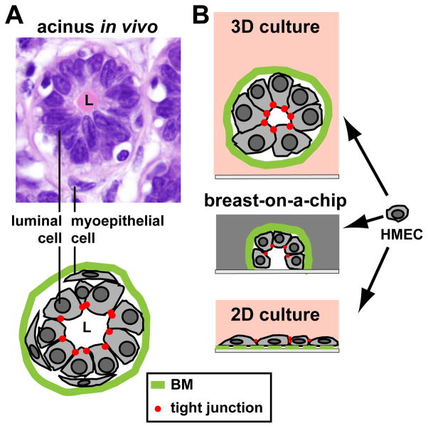
Figure 2.
Cell culture models for the breast epithelium. A Hematoxylin & Eosine staining of normal breast tissue showing the organization of luminal (inner layer) and myoepithelial (outer later) cells in the smallest structural and functional unit (acinus) of the breast glandular epithelium. On the drawing, the basement membrane (BM) and apical tight junctions are represented. B Cultures of human mammary epithelial cells (HMECs): flat cell monolayers (2D culture) do not recreate the organization of the glandular and ductal epithelia. In contrast, three-dimensional (3D) cultures of HMECs in compliant hydrogels yield growth-arrested spheroid structures with basal polarity (cell-BM contacts) and apical polarity (apical tight junctions) mimicking glandular structures or acini. When placed on chip devices resembling a ductal system (breast-on-a-chip), cells divide and spread alongside the walls of the hemichannel, leading to a growth-arrested basoapically polarized monolayer of cells.

