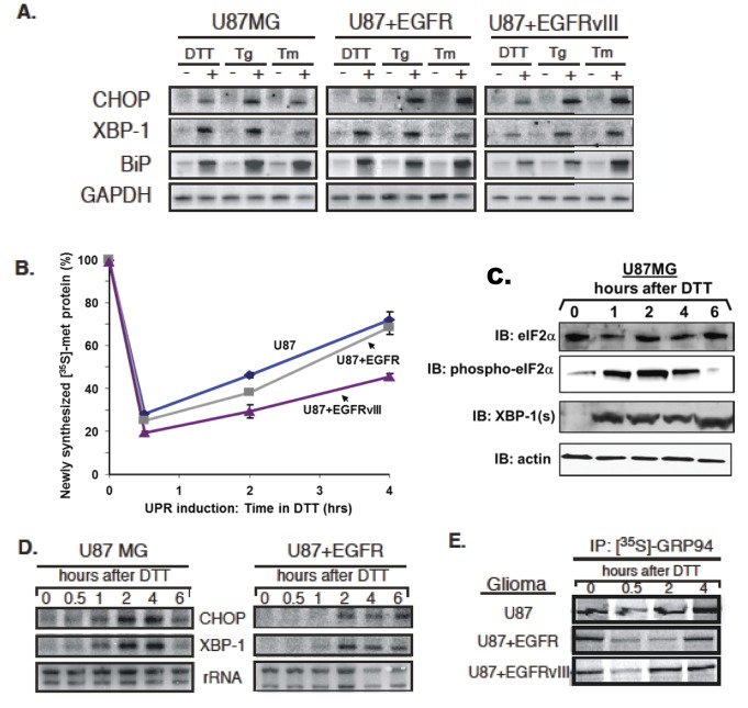
Figure 4. Human glioma cells respond to persistent ER stress with UPR induction and rapid recovery of protein synthesis.
Human glioma tissue culture models examined include: U87MG, U87+EGFR, and U87+EGFRvIII, from U87 parent lines stably transfected with a constitutively-active oncogenic EGFR and the extracellulary truncated EGFR variant III. (A) Northern blot analysis of human glioma tissue culture cells after a 4-hour treatment with the following pharmacological inducers of the UPR: 1mM dithiothreitol (DTT), 0.5 µM thapsigarin (Tg), or 2.5µg/ml tunicamycin(TM). Blots were probed for message levels of CHOP, XBP-1, GRP78/BiP, and GAPDH. (B) Levels of total newly-synthesized protein from 0–4 hours after a 1mM DTT treatment were assayed as TCA precipitable [35S]-methionine-labeled protein. Protein synthesis rapidly declined and then rapidly recovered despite the presence of reducing agent in the culture. (C) The time course of eIF2α phosphorylation (“phospho- eIF2α”) was followed by immunoblotting during DTT treatment of U87MG cells, as was induction of spliced XBP-1 (“XBP-1(s)”. The actin loading control blot is a replicate blot. Following a 0-6 hour 1mM DTT treatment in U87 cell culture, (D) RNA was analyzed by Northern blot in a kinetic analysis of CHOP and XBP-1 mRNA. (E) Glioma cell cultures were labeled with [35S]-methionine during a 0-4 hour 1mM DTT treatment. GRP94 was immunoprecipitated to detect newly-synthesized protein.

