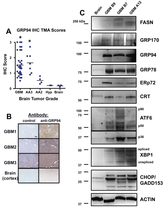
Figure 8. Immunohistochemistry and Western blots of patient brain tumors reveal high expression of UPR-related proteins.
A Cybrdi “brain glioblastoma” tissue microarray (TMA) was probed for GRP94 by immunohistochemistry (IHC); (A) Scores were derived as describe in Materials and Methods, and show that high grade tumors such as glioblastoma multiforme (GBM, WHO grade IV) and anaplastic astrocytomas (WHO grade III—AA3) express significantly higher levels of GRP94 than do lower grade tumors (grade II anaplastic astrocytomas, AA2), anaplastic hyperplasia (Hyp) or normal brain (*, p < 0.05 by ANOVA comparing high grade gliomas vs the rest of the samples). Examples of the IHC staining are shown for 3 GBMs and normal brain (B). (C) Grade IV (GBM) tumor lysates (3 different tumors, not the same as those in B) and normal brain (cortex) lysates were separated by SDS-PAGE and electroblotted for Western blotting. Blots were probed with the antibodies listed as in Figures 2 and 3; “FASN” = Fatty Acid Synthase; “p90/p50/p36” = full length and cleaved forms of ATF6 “spliced/unspliced” = spliced or unspliced protein product of XBP1. Molecular weight markers are listed at left. Actin blot shown as loading control was a replicate for GRP78 and CRT.

