
Fig. 5.
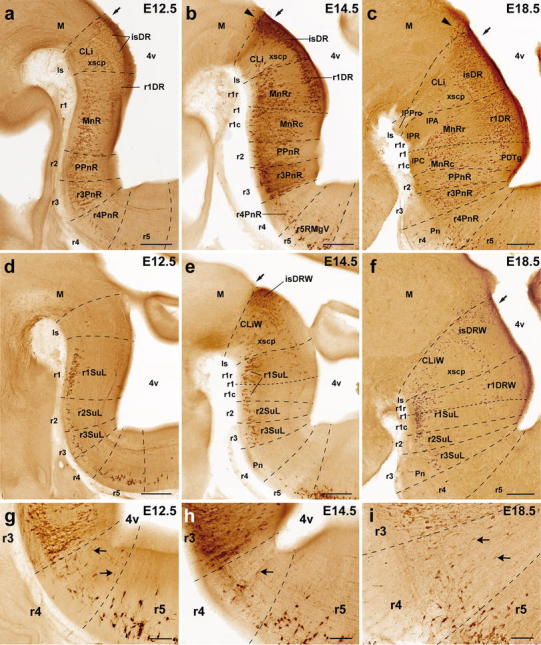
Segmental mapping of the rostral raphe cluster during embryonic development. 5-HT immunoreactive neurons observed in sagittal sections of mouse brains at E12.5 (a, d), E14.5 (b, e), and E18.5 (c, f), with superposed tracing of postulated interrhombomeric boundaries (dashed lines), and our tentative identification of the nuclear primordia (Table 1). Each set of three images read from left to right (e.g., a–c) represents a temporal sequence at a given section plane. Arrows mark the midbrain–hindbrain boundary. a–f Rostral cluster at paramedian section level. d–f Rostral cluster at a more lateral level. g–i Details at higher magnification of the paramedian pontine region of Fig. 5a–c, respectively. Note some 5-HT immunopositive cells are always present in r4, mainly in its superficial stratum, though cells with weaker immunoreaction are also observed in the intermediate stratum (arrows in g–i). For abbreviations see "List of abbreviations". Scale bar 250 μm in a–f, 100 μm in g–i