Fig. 2.
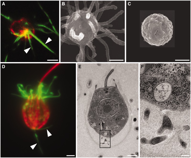
Filopodia-like structures in close relatives of metazoans. (A) Capsaspora owczarzaki filopodiated cells bear multiple long bundles of actin microfilaments, as revealed by staining with phalloidin (green). The cell periphery is revealed by staining with antibodies against beta-tubulin (red). SEM shows the presence of multiple long filopodia-like structures in C. owczarzaki filopodiated cells (B), but not in nonfilopodiated, cystic cells (C). (D) In Salpingoeca rosetta, attached cells bear actin microfilaments in the apical collar of microvilli and in basally positioned long cellular protrusions that resemble filopodia. Salpingoeca rosetta cells were stained with phalloidin (green) and antibodies against beta-tubulin (red). (E) TEM of thin sections through a choanoflagellate shows the presence of basally positioned cellular processes (indicated with black rectangle), shown in higher magnification in (F). Scale bars (A–E: 1 µM, F: 200 nm).

