Fig. 3.

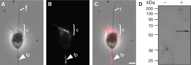
Subcellular localization of Fascin in Salpingoeca rosetta. (A) Phase contrast microscopy shows the morphology of a fixed S. rosetta cell. (B, C) Immunolocalization studies reveal that Fascin localizes to a basal filopodia-like structure (fp) and to the apical actin filled collar (c). (D) Western blot analysis shows that S. rosetta cell lysate probed with Fascin antibodies detect a single band of approximately 55 kDa (+). No signal was detected when primary Fascin antibody was omitted (−). f, flagellum; c, microvilli collar; fp, filopodia. Scale bar: 1 µM.
