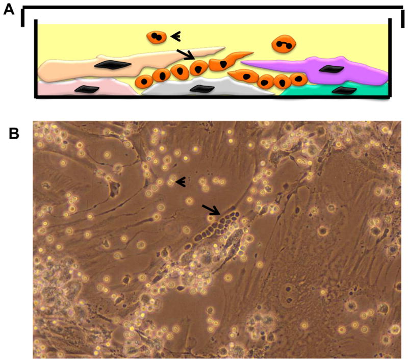
Figure 4. Primary Long Term Cultures (LTCs or Dexter Cultures).

A: Cartoon depicting vertical layer of an LTC with stromal and hematopoietic elements.
B: Phase contrast micrograph of an LTC.
Long arrows in both panels depict the cobble stone areas (CSAs) comprised of primitive hematopoietic precursors trapped within the stromal layers and appear as “phase-dark” cells resembling a cobble-stone. Mature myeloid cells are released into the supernatant when the more primitive precursor cells in the CSAs divide and mature ( short arrow in both panels) and appear as “phase-light cells” in phase-contrast micrographs.
