
Figure 7.
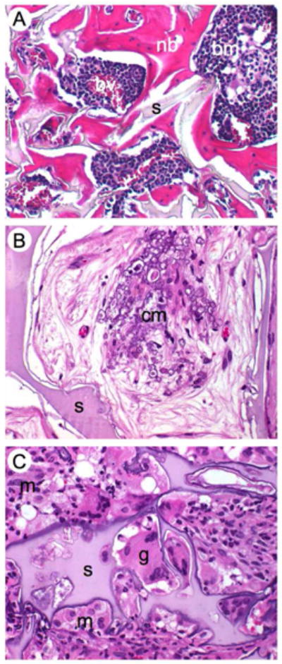
Histological details of mouse calvarial defects 8 weeks after surgery and stained with H&E. (A) Newly formed bone around the silk fibroin scaffold decorated with RGD sequences showing normal bone morphology, bone marrow and vascularization. (B) Clusters of beginning mineralization in the pore void, (C) silk with mineralizing edges surrounded by foreign body giant cells, neutrophils and macrophages that start degrading the silk-RGD. nb = newly formed bone, s = silk, bm = bone marrow, bv = blood vessels, cm = clusters of mineralization, g = giant cell, m = macrophage, n = neutrophil. Magnification 40x