Figure 1.

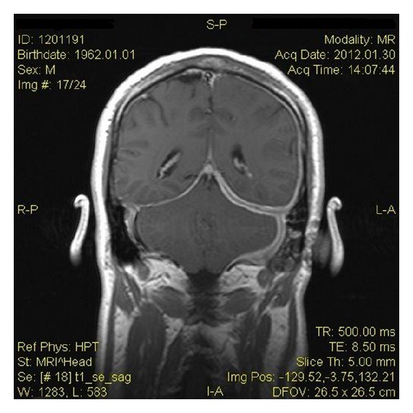
Gadolinium-enhanced coronal T1-weighted MRI scan showing pachymeningeal enhancement over the left temporal cerebral convexity, the left cerebellar convexity, and the left side of tentorium cerebelli. Also, there is left mastoiditis.

Gadolinium-enhanced coronal T1-weighted MRI scan showing pachymeningeal enhancement over the left temporal cerebral convexity, the left cerebellar convexity, and the left side of tentorium cerebelli. Also, there is left mastoiditis.