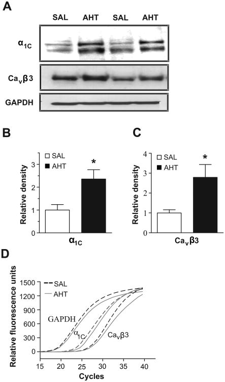
Figure 1.
A. WB showing adjacent lanes loaded with 40 μg of MA protein lysate pooled from either two SAL or AHT mice and probed with anti- α1C and Cavβ3. GAPDH was a loading control. B, C. Densitometric analyses of α1C and Cavβ3 immunoreactivity (n=6; * = p<0.05). D. qRT-PCR amplification corresponding to α1C, Cavβ3 and GAPDH transcripts averaged from 4 MA samples from SAL and AHT mice.

