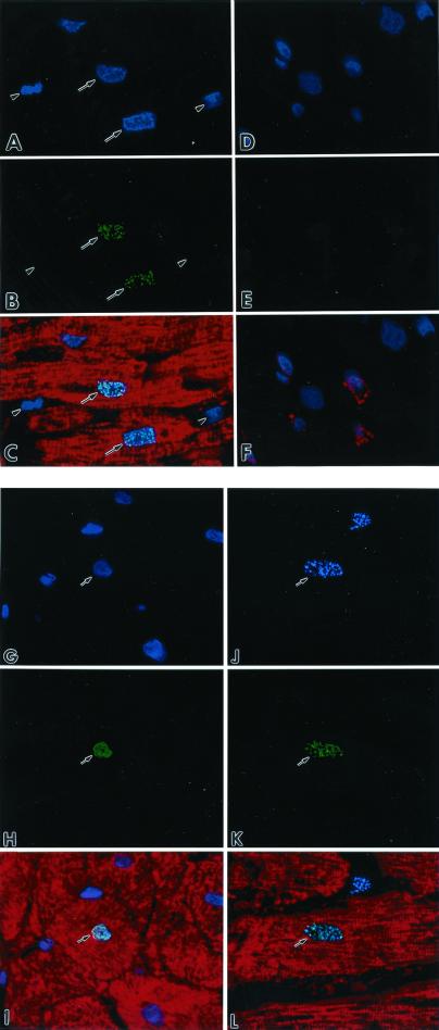
Figure 2.
(A and D) Nuclei by the blue fluorescence of PI. (B) Green fluorescence telomerase staining (arrows) of nuclei. (E) The absence of telomerase labeling. Red fluorescence cardiac myosin staining of myocyte cytoplasm (C) and factor VIII labeling of endothelial cells (F). Bright fluorescence in C corresponds to the combination of PI and telomerase staining of nuclei. Arrowheads, interstitial nuclei negative for telomerase. (G) Nuclei by the blue fluorescence of PI. (H) Green fluorescence Ki67 staining of a nucleus. (I) Myocyte cytoplasm by the red fluorescence of cardiac myosin. Bright fluorescence (I) corresponds to the combination of PI and Ki67 localization in a myocyte nucleus. (J) Blue fluorescence Ki67 labeling of a nucleus. (K) Green fluorescence telomerase staining in the same nucleus. (L) The myocyte cytoplasm by the red fluorescence of cardiac myosin. Bright fluorescence (L) corresponds to the colocalization of Ki67 and telomerase in the myocyte nucleus. Confocal microscopy, ×1,000.

