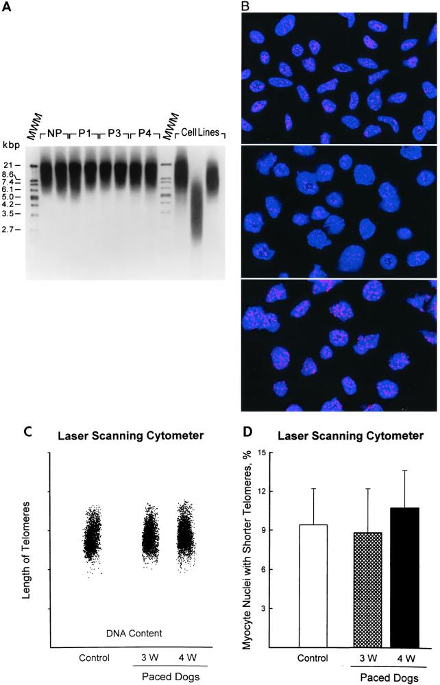
Figure 4.
(A) TRF length in myocytes from nonpaced (NP), P1, P3, and P4 dogs. Immortal cell lines used for comparison have, from left to right, a mean TRF of 10.2, 3.9, and 7 kbp, respectively. MWM, molecular weight marker. (B) Myocyte nuclei (Top) and lymphoma cells with short (Middle) and long (Bottom) telomeres were stained by in situ hybridization with a PNA probe specific for telomeric sequence. Nuclei are illustrated by the blue fluorescence of PI, and the red fluorescent dots correspond to individual telomeres. Confocal microscopy, ×1,000. (C) Bivariate distribution of DNA content and telomere length in myocyte nuclei from the left ventricle of nonpaced and paced dog hearts. (D) Percentages of myocyte nuclei with shorter telomeres. Results are mean ± SD; n = 6 in each group of dogs.

