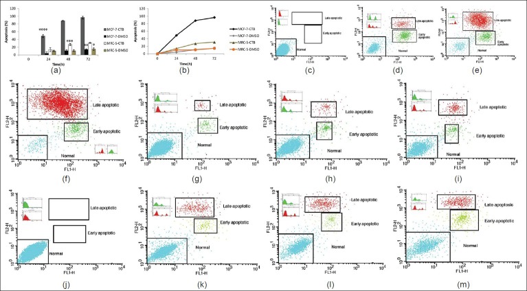
Figure 2.
CTB induces apoptosis in cancer cells lines (MCF-7) but not fibroblasts (MRC-5). Relative levels of apoptotic cells in MCF-7 cancer cell lines and cultured fibroblasts (MRC-5) treated with 85.43 μM CTB for different times. Cells incubated with the vehicle (DMSO) were used as a control. (a, b). The percentage of apoptotic cells was measured using the AnnexinV FITC and PI assay as described in Materials and Methods. ****P <0.001 vs. all other groups MCF-7 cells treated with CTB. ***P<0.05 vs. all other groups MRC-5 cells. **P< 0.05 vs. all other groups MCF-7 cells incubated with the vehicle (DMSO) were used as a control. *P< 0.05 vs. 48 and 72 hours groups MRC-5 cells incubated with the vehicle (DMSO) were used as control. (c-m) MCF-7 and MRC-5 cells were treated with 85.43 μM of CTB, and apoptosis was measured by flow cytometry following AnnexinV (FL1-H) and PI (FL2-H) staining. Cells that are AnnexinV-positive and propidium iodide negative are in early apoptosis, as phosphatidyl serine (PS) translocation has occurred, although the plasma membrane remains intact. Cells that are positive for both AnnexinV and PI either are in the late stages of apoptosis or are already dead, as PS translocation has occurred, and the loss of plasma membrane integrity is visible

