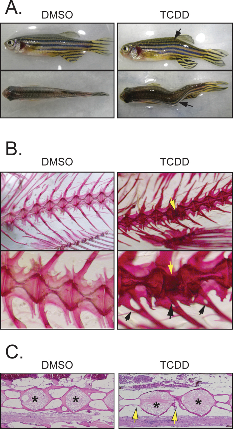
Fig. 2.
Sublethal exposure causes abnormalities in the F0 axial skeleton. Zebrafish larvae were exposed to TCDD at 50 pg/ml (6.4nM) at 3 and 7 wpf and collected at 1 year of age as described in the Materials and Methods section. (A) Photographs of control and treated examples showing the lateral and dorsal views. The black arrows indicate a kink in the axial skeleton. (B) Alizarin red staining showing control and treated axial skeletons. The bottom row shows higher magnification of the images in the top row. The open arrows point to a defect in one of the vertebrae, whereas the black arrows point to malformed vertebral rays. (C) H&E-stained sagittal sections from control and treated fish. Arrows point to defects in the vertebral bodies; asterisks indicate region of the growth plate.

