
Figure 7. In vivo IL-12p40 blockade restores optimal T cell tuning in MPF-housed hosts.
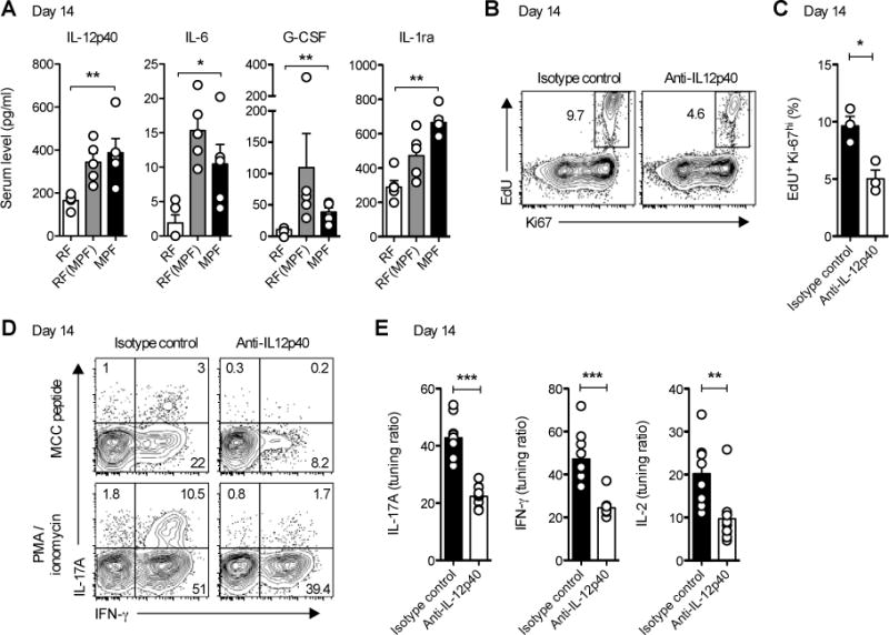
(A) IL-12p40, IL-6, G-CSF and IL-1Ra levels in sera of mPCC, Cd3e−/− hosts 14 days post naïve 5C.C7 T cell transfer. Data (mean±SEM) pooled from 2 independent experiments (n=5 mice per group).
(B–E) Starting at day 7, MPF-housed mPCC, Cd3e−/− mice were further injected twice daily i.p. with anti-IL12p40 or a corresponding isotype control mAb. At day 14, mice were injected with EdU and sacrificed 1hr later. (B) Representative EdU and Ki67 expression profiles and (C) frequency of EdU+Ki-67hi cells in live, CD4+Vβ3+ T cells isolated from mLN (n=3 mice per group, mean±SEM). * P<0.05, student t test. (D) Representative IL-17A and IFN-γ expression profiles and (E) tuning ratio for IL-17A (left), IFN-γ (middle) and IL-2 (right) production in live, CD4+Vβ3+ T cells isolated from mLN at day 14 and restimulated for 3h with PMA and ionomycin or 3μM MCC peptide. Data (mean±SEM) pooled from 4 independent experiments (n=9–10 mice per group). See also Figure S6.