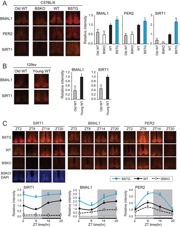
Figure 4. SIRT1 up-regulates circadian proteins in the SCN and declines with aging.
(A) Typical immunohistochemical staining results and relative signal intensities for BMAL1, PER2 and SIRT1 proteins in the SCN. Sections were prepared from C57BL/6 background mice that were sacrificed at ZT4 (n≥3 mice/genotype). Aged WT mice (22 months) are compared to young BSKO, WT and BSTG mice (5 months). Signal intensities of protein levels were quantitated and are shown relative to wild type (≥ 6 sections). (B) Typical immunohistochemical staining images and relative signal intensities of BMAL1 and SIRT1 proteins in the SCN. Sections were prepared from aged (21 month) or young (4 month) 129 sv background mice that were sacrificed at ZT4 (n≥3). Signal intensities of protein levels were quantitated and are shown relative to young animals (≥ 6 sections). Values represent the mean ± standard deviation. *P<0.05; **P<0.01; t test. (C) Temporal analysis of BMAL1, PER2 and SIRT1 proteins in the SCN. SCN sections were prepared from 3 month-old mice that were sacrificed at indicated time (n≥3 mice/genotype/time point). DAPI stained images were shown to indicate SCN in the BSKO sections. Signal intensities of protein levels were quantitated and are shown relative to wild type (≥ 6 sections). Values represent the mean ± standard deviation. *P<0.05; **P<0.01; t test.

