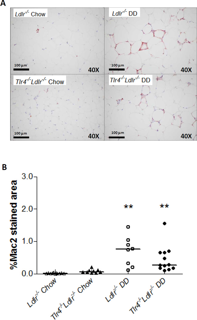
Figure 3. Intra-abdominal adipose tissue macrophage accumulation is not reduced in Tlr4−/−Ldlr−/− mice.
Representative photomicrographs of epididymal adipose tissue stained with a macrophage-specific antibody Mac2 (1:2000 dilution, red), 40×magnification, n=10–13 per group (A), and quantification of Mac2 staining (B); (**P<0.01 vs DD or chow groups).

