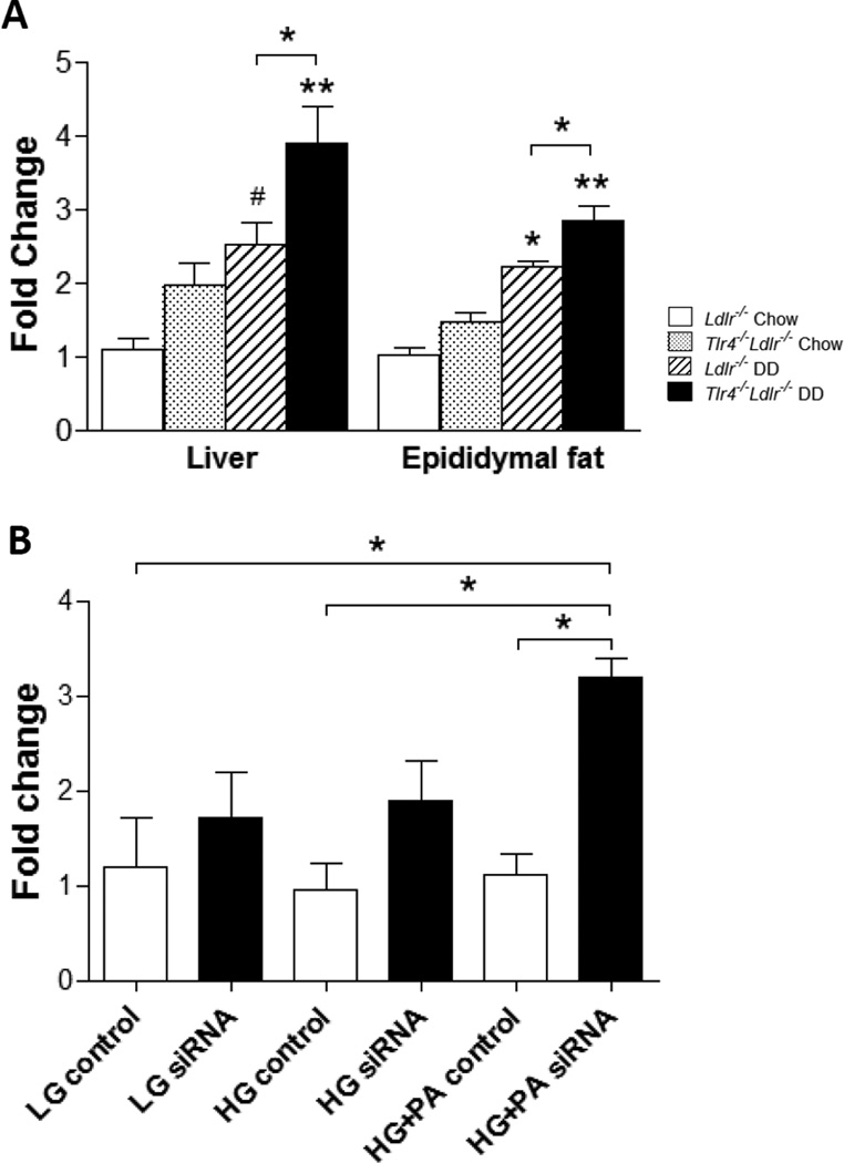
Figure 5. TLR2 expression is increased in DD-fed TLR4 deficient mice and TLR4-silenced differentiated 3T3-L1 adipocytes treated with FFA.
Tlr2 mRNA expression in liver and epididymal adipose tissue from TLR4-deficient mice (A); (n=10–15, *P<0.05, **P<0.01 vs DD or chow groups). Tlr4 mRNA expression in differentiated 3T3-L1 cells (B). 3T3-L1 adipocytes were transfected with a siRNA specific for TLR4 or a scrambled siRNA as control. 24 hours later, the cells were exposed to palmitate (PA, 250 µmol/L) in 5 (LG) and 25 mmol/l (HG) for 7 days with daily medium changes (*P<0.05 as tested by one way ANOVA compared with LG control). Tlr4−/−Ldlr−/− mice fed DD (black bars) and chow (grey bars), Ldlr−/− control mice fed DD (hatched bars) and chow (open bars).

