Figure 5.
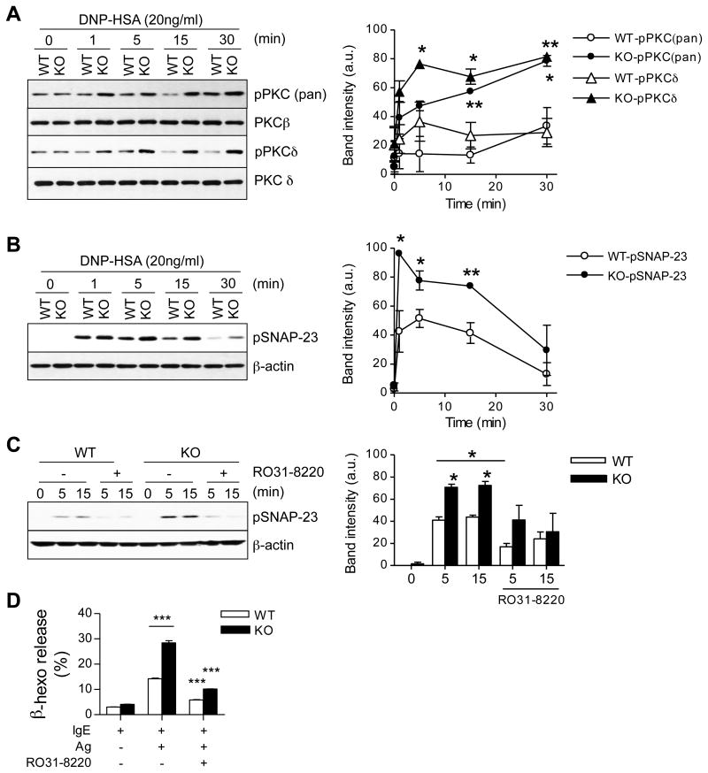
Increased phosphorylation of PKCs and SNAP-23 in lipin1-deficient mast cells. (A, B) Cell lysates were generated and subjected to immunoblotting analysis as described in Fig. 4 by using anti-phospho-pan-PKC, anti-phospho-PKCδ (A) and anti-phospho-SNAP-23 (Ser95) (B) antibodies. Total PKCβ/δ and β-actin were used as the loading controls. Graphs are mean ± SEM presentation of band intensities determined by densitometry. (C) Inhibition of SNAP-23 phosphorylation in WT and lipin1-KO BMMCs is dependent on PKC activity. WT and lipin1-KO BMMCs were incubated with or without 10 μM RO31-8220 for 30 min and then left unstimulated or were stimulated with DNP-HSA, followed by immunoblotting analysis. Bar Graph is mean ± SEM presentation of band intensities determined by densitometry. (D) Enhanced degranulation by lipin1-KO BMMCs is dependent on PKC activity. IgE-sensitized BMMCs were treated with RO31-8220 (10 μM) for 30 min, followed by in vitro mast cell degranulation assay, as indicated in Fig. 2. Data shown are representative from three experiments. * p<0.05, ** p<0.01 and ***, p<0.001 by Student t-test.

