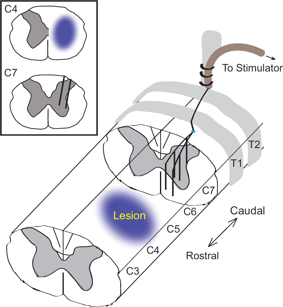
Figure 2.
Diagram showing cervical contusion injury and placement of intraspinal stimulating electrodes. The five 30 µm platinum-iridium electrodes are routed along the surface of the spinal cord before passing through a small incision in the dura and penetrating to a depth of 1.2–1.8 mm within the spinal cord. The dura is then sutured closed and sealed with a microdrop of cyanoacrylate, and the implant is secured to T2 with sutures. The return electrode is placed above the spinal cord (not shown).

