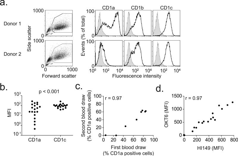
Figure 1. Deficiency of CD1a on human dendritic cells.
(a) Viable monocyte-derived dendritic cells, identified by high forward and side scatter profiles, were stained with fluorescently-conjugated antibodies against CD1a, CD1b, and CD1c (dark lines) as well as isotype control antibody (shaded histogram). Shown are representative plots from two donors. (b) Median fluorescence intensity (MFI) of CD1a and CD1c is shown for 19 healthy blood donors. In four donors, CD1a MFI is less than 10. P-value reflects Bartlett's test for non-homogeneity of variances. (c) Simple linear correlation between CD1a staining results of sequential blood draws for eight subjects. Data are represented as % positive cells rather than MFI to adjust for temporal variation in flow cytometry calibration. (d) Simple linear correlation of staining between two antibodies (OKT6 and HI149) that bind CD1a.

