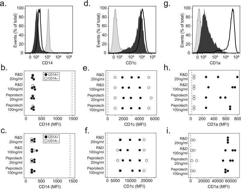
Figure 2. CD1a-deficiency does not reverse with increased IL-4 concentrations or extended time in culture.
Human monocytes were cultured with IL-4 sourced from either Peprotech or R&D Systems at a concentration of either 20ng/ml or 100ng/ml for either three or six days. Cells were harvested and stained for flow cytometry with fluorescently conjugated antibodies against CD14, CD1a, or CD1c. (a) Shown are expression profiles from unstimulated monocytes (shaded histogram) or cells cultured for six days from a CD1a-sufficient donor (dark line), and a CD1a-deficient donor (dark histogram) for CD14. Results from five subjects are combined and shown as the median fluorescence intensity (MFI) of CD14 after (b) three days of culture or (c) six days of culture. (d) Representative CD1c expression from six day cultured cells or combined data showing CD1c MFI after (e) three days or (f) six days of culture. (g) Representative CD1a expression from six day cultured cells or combined data for CD1a after (h) three days or (i) six days of culture. Expression levels on unstimulated monocytes (dotted line) are shown for comparison. All data are representative of two independent experiments.

