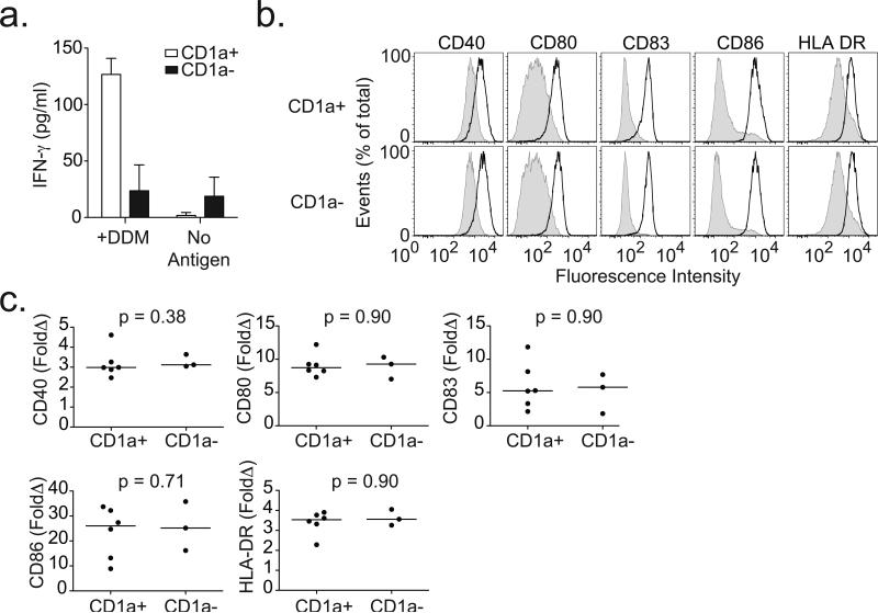
Figure 4. CD1a-deficient dendritic cells fail to present antigen to T cells but are able to induce expression of co-stimulatory molecules.
(a) CD1a-sufficient (CD1a+) or CD1a-deficient (CD1a-) dendritic cells were co-incubated with T cell clone CD8-2 in the presence or absence of mycobacterial lipopeptide antigen dideoxymycobactin (DDM). IFN-γ levels were measured in overnight supernatants by ELISA for one donor per group. Shown are mean and standard deviation for triplicate measurements. (b) Shown are expression profiles for cells derived from two healthy blood donors and treated with media (shaded) or LPS (dark line) overnight and then stained with antibodies against co-stimulatory molecules and HLA-DR. (c) Results from nine subjects are combined and shown as fold change (LPS/Media) in median fluorescence intensity (MFI) for CD40, CD80, CD83, CD86, and HLA-DR. All data are representative of two or three independent experiments.

