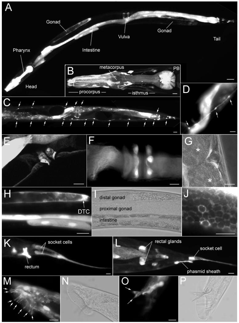
Figure 3. T27A3.1 is expressed in many cell types.
(A) Micrograph showing expression of T27A3.1pro::gfp in an hermaphrodite sfEx27 adult worm. GFP is visible in cells in various organs including the pharynx, the intestine, the gonads, as well as cells in the vulval and tail regions.
(B) Micrographs of T27A3.1pro::gfp expression in the pharynx of an hermaphrodite sfEx27 adult worm. GFP positive cells are located within the posterior bulb, in what appears to be muscle cells. GFP is also visible in cells around the pharyngeal bulb as well as in processes along the bulb (arrows) and in the nerve ring (arrowheads).
(C) Micrographs showing GFP expression in the seam cells (arrows) along the body of an hermaphrodite sfEx27 adult worm.
(D) Higher magnification of GFP expression in the seam cells (arrows).
(E) Lateral view of the vulval region of an hermaphrodite sfEx27 adult worm showing GFP in cells surrounding the vulva (arrowheads).
(F) Ventral view of the vulva region showing GFP in cells surrounding the vulva (arrowheads).
(G) Lateral view of the vulval region showing the position of the GFP positive cells relative to embryos (*).
(H) Micrographs of T27A3.1pro::gfp expression in the gonads of an hermaphrodite sfEx27 adult worm. GFP is visible in the DTC cell as well as in the intestine.
(I) Same as H but shown in phase contrast mode.
(J) High magnification of fish-net like GFP expression in the distal sheath cell pair 1 of the gonads.
(K) Micrographs of the tail region of a T2A3.1pro::gfp adult sfEx27 hermaphrodite. GFP is visible in the rectum and in socket cells.
(L) In the tail region, GFP is also found in the rectal glands and phasmid sheath cells.
(M) Micrograph of the tail region of a T2A3.1pro::gfp adult male sfEx27 showing GFP expression in the rays (arrows).
(N) Same as H in phase contrast mode.
(O) In the male tail, GFP is also visible in the spicules (arrows).
(P) Same as H in phase contrast mode. Scale bars: A: 100 μm; B-D: 20 μm; E-J: 10 μm; K: 40 μm; L: 20 μm; M-P: 10 μm.

