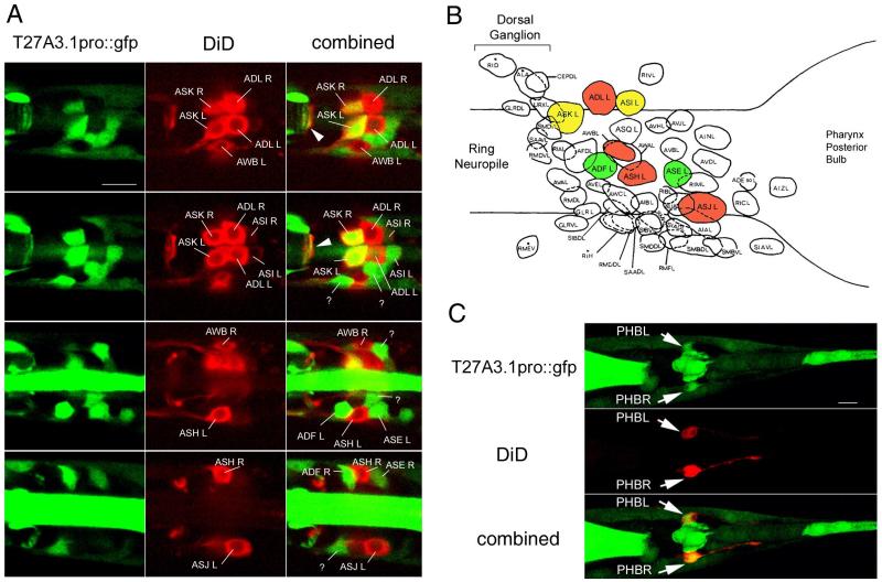
Figure 5. T27A3.1 is expressed in some head and tail chemosensory neurons.
(A) Confocal micrographs of the head region of an adult sfEx27 stained with DiD. Each row of images represents a different dorsal to ventral view to allow us to visualize all of the DiD labeled neurons. DiD staining is shown in red and GFP in green. In the head, DiD is known to back-fill 6 pairs of amphid neurons including ASK, ASI, ADL, AWB, ASH and ASJ neurons which can be identified based on their respective positions. T27A3.1pro::gfp is expressed in the ASK and ASI pairs of chemosensory neurons. In addition to being present in some amphid neurons, GFP was also present in ADF and ASE chemosensory neurons. Arrows indicate overlapping GFP and DiD in neuronal processes in the nerve ring.
(B) Schematic showing the position of the various neurons in the posterior ganglion modified from Wormatlas. T27A3.1pro::gfp expressing neurons are identified in green, DiD back-filled chemosensory neurons are in red, overlapping is shown in yellow.
(C) Confocal micrographs of the tail region of an adult sfEx27 hermaphrodite stained with DiD. In the tail region, DiD back-fills 2 pairs of phasmid neurons. The most anterior pair of neurons (arrows) corresponding to PHBL and PHBR shows overlapping GFP and DiD. Scale bars: A and C: 10 μm.

