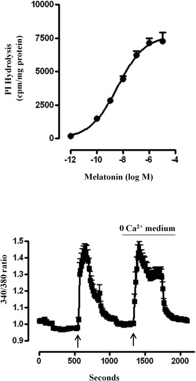
Figure 3. Stimulation of PLC-β activity and release of Ca2+ by melatonin.
(A) Phosphoinositide-specific (PI) hydrolysis (PLC-β activity) in response to melatonin was measured in dispersed muscle cells labeled with myo-[3H]inositol. Freshly dispersed muscle cells were treated for 60 s with different concentrations of melatonin and PLC-β activity was measured as increase in water-soluble [3H]inositol formation. The results are expressed as [3H]inositol phosphate formation in counts per minute (cpm) per mg protein above basal levels (basal: 642±99 cpm/mg protein). Values are means±SEM of 4 experiments. (B) Isolated smooth cells were loaded with 5 μM fura-2 and treated with 1 μM melatonin in the absence of extracellular Ca2+. The cells were alternately excited at 380 nm and 340 nm. The background and autofluorescence were corrected from images of a cell without the fura 2. Results are expressed as 340/380 ratio and an increase in ratio reflects an increase in cytosolic Ca2+. The figure shows results obtained from 38 cells.

