
Fig. 1.
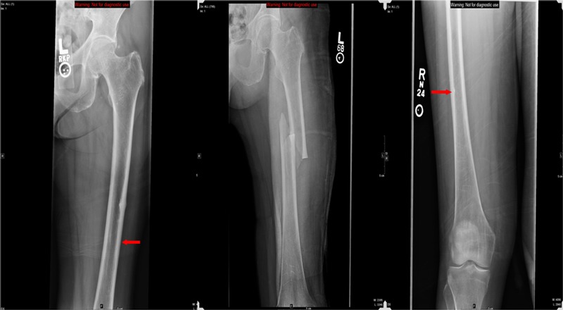
Radiographic appearance of an atypical femoral fracture in a sixty-three-year-old woman who had been on bisphosphonate therapy for seven years prior to developing left thigh pain. The left panel is an initial anteroposterior radiograph of the left femur showing focal cortical thickening along the lateral proximal femoral diaphysis that represents an incomplete fracture (arrow). The central panel is an anteroposterior radiograph of the femur taken two days later showing a displaced fracture centered at the prior site of cortical thickening even though the patient had been placed on protected weight-bearing and did not sustain a trauma during the intervening time. The right panel is an anteroposterior radiograph of the right femur of the same patient, made after she had begun experiencing milder right thigh pain, demonstrating subtle cortical thickening along the lateral proximal aspect of the right femur that also represents a developing insufficiency fracture (arrow).