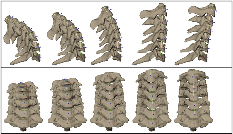
Fig. 2.

Three-dimensional bone models at five instants of the flexion cycle from a representative control subject. Sagittal views are above, and coronal views are below. Intervertebral kinematics were determined for each pair of adjacent vertebrae (superior bone motion relative to immediately inferior bone) with use of coordinate systems embedded within each vertebral body.
