Fig. 3.

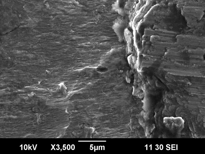
Fig. 3 Scanning electron microscope image (×3500) demonstrating fretting and corrosion (left and center) as well as a thick deposit of fretting-corrosion debris (right) on the lateral aspect of a damaged neck-body junction that had been in vivo for approximately twenty months (Case 1). Similar findings were seen in all ten retrieved implants.
