Fig. 5.

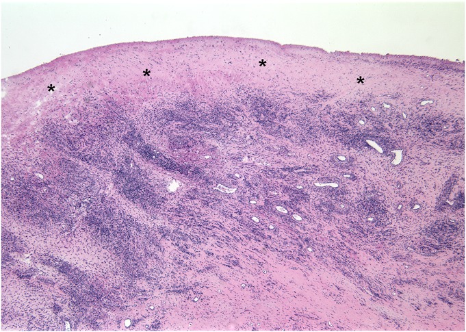
Fig. 5 Histological surgical specimen obtained 12.6 months following hip arthroplasty (Case 4), demonstrating abundant diffuse and perivascular lymphocytes in the joint pseudocapsule. The synovial lining is absent, and a layer of organizing fibrin exudate (*) covers the inner surface of the capsule (hematoxylin and eosin stain, ×25 magnification).
