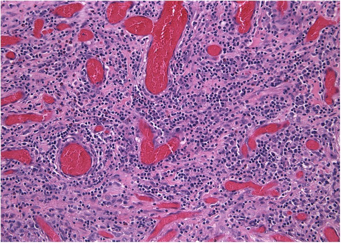
Fig. 7.

Histological specimen removed during revision surgery (Case 1). The majority of the specimen was necrotic, but viable areas of pseudocapsule showed chronic inflammation dominated by diffuse lymphocytes and scattered eosinophils (hematoxylin and eosin stain, ×200 magnification).
