
Figure 1. Quantitative proteomic analysis of FFPE melanoma.
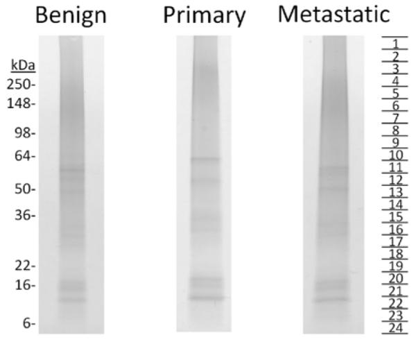
Melanocytes from benign FFPE skin biopsies (25 total) and melanoma cells from primary (12 total) and metastatic melanoma (24 total) FFPE biopsies were isolated by needle dissection. Protein extracts were resolved by SDS-PAGE, visualized by Coomassie staining, excised as 24 bands per lane, and subjected to in-gel trypsin digestion. Tryptic peptides were analyzed by LC-MS/MS and relative protein levels were determined by spectral counting [9].