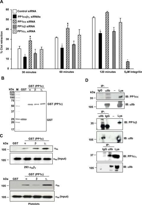
Figure 4. Effect of depleting the PP1c isoforms on fibrin clot retraction.
(A) The decreased fibrin clot retraction of PP1c (α, β, γ1) depleted cells compared to the control siRNA treated cells was significant at *p=0.03, *p=0.01, *p=0.001 for 30, 60 and 120 minutes, respectively. The increased fibrin clot retraction of PP1cα depleted cells compared to the control siRNA treated cells was significant at ◆p=0.05, ◆p=0.04, p=0.07 for 30, 60 and 120 minutes, respectively. The decreased fibrin clot retraction of PP1cβ depleted cells compared to the control siRNA treated cells was significant at †p=04, †p=.0.03, †p=0.005 for 30, 60 and 120 minutes, respectively. Integrilin (5 μM) blocked the clot retraction of control siRNA cells at 120 minutes *p=0.003. n=3. (B) PP1c α, β and γ1 GST proteins separated on a SDS-PAGE gel were detected by Coomassie Blue staining. M is Molecular weight marker. (C). αIIb in the lysate from the 293 αIIbβ3 cells and platelets interact with PP1cα–GST, PP1cβ-GST, PP1cγ1–GST but not GST. Comparable levels of αIIb in lysate (input) was used for pull down studies. n=2-3. (D) 293 αIIbβ3 cell lysate was immunoprecipitated (IP) with IgG (control) or αIIb and immunoblotted (IB) with anti- PP1cα, PP1cβ, PP1cγ1 and αIIb. Lys is cell lysate. n=2.

