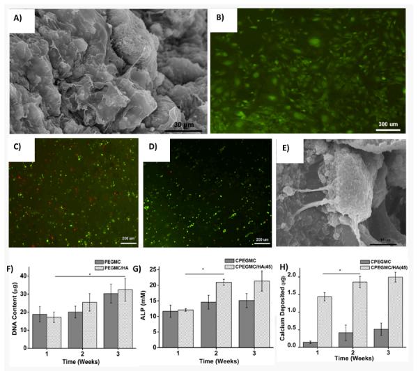
Fig. 6.

hFOBs attachment on the surface of CPEGMC/HA (45) films 48 hours post-seeding. A) SEM image of hFOB-seeded PEGMC/HA(45) composite and B) Microphotograph of hFOBs on CPEGMC/HA(45) composite stained with CFDA-SE flourecent dye. hFOBs encapsulated in CPEGMC/HA(45) composites. C) Live/Dead stain of hFOBs encapsulated in CPEGMC/HA(45) after 1 day culture; D)) Live/Dead stain of hFOBs encapsulated in CPEGMC/HA(45) after 21 day culture; Live cells are green fluorescent and dead cells are red fluorescent; E) Crossectional SEM image of hFOBs encapsulated within the matrix; F) DNA content assay; G) alkaline phosphatase (ALP) assay; and H) Calcium assay. In F-H, hFOBs were encapsulated in CPEGMC and CPEGMC/HA(45).
