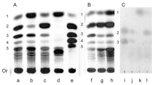
Fig. 5. TLC analysis of β-eliminated O-glycans of mntΔ mutant strains.
A, cell walls were labeled by growing whole exponentially growing yeast cells in d-[2-3H]mannose and removing O-glycans by β-elimination then separating them by TLC. Labeled products then visualized by autofluorography were from: lane a, wild-type C. albicans CAF2-1; lane b, Camnt1Δ; lane c, Camnt2Δ; lane d, Camnt1-Camnt2Δ; and lane e, wild-type S. cerevisiae serving as a labeled Man1-Man5 control. B, control strains with: lane f, the parent CAI4+CIp10; lane g, Camnt1-Camnt2Δ + CIp10-MNT1; and lane h, Camnt1-Camnt2Δ + CIp10-MNT2. C, confirmation of α-1,2 linkages in O-glycans by mannosidase digestion. Standards (lane i), and untreated (lane j) purified cell walls of CAF-2 were used and were extracted by reductive β-elimination. Cell walls were then digested with Jack Bean mannosidase (lane k) and A. satoi mannosidase (lane l). Positions of Man-OH, Man1, Man2, and Man3 are shown. Or, origin.

